How Cells Communicate During Fight or Flight

When our senses perceive an environmental stress such as danger or a threat, cells in the nervous and endocrine systems work closely together to prepare the body for action. Often referred to as the fight or flight or stress response, this remarkable example of cell communication elicits instantaneous and simultaneous responses throughout the body.

Initiating the Response

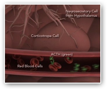
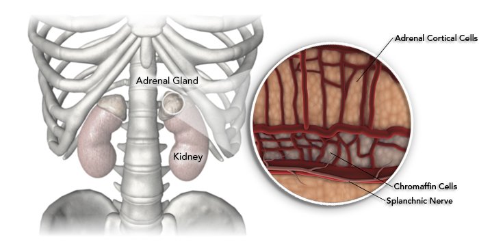
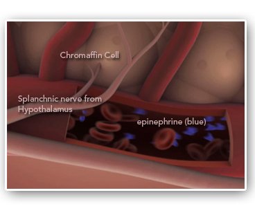
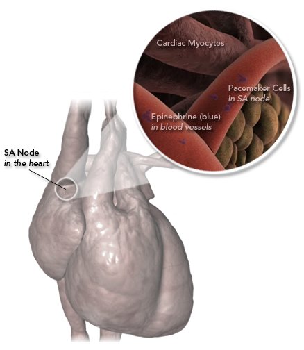
Sensory nerve cells pass the perception of a threat, or stress, from the environment to the hypothalamus in the brain. Neurosecretory cells in the hypothalamus transmit a signal to the pituitary gland inciting cells there to release a chemical messenger into the bloodstream. Simultaneously, the hypothalamus transmits a nerve signal down the spinal cord. Both the chemical messenger and nerve impulse will travel to the same destination, the adrenal gland.

Pituitary gland

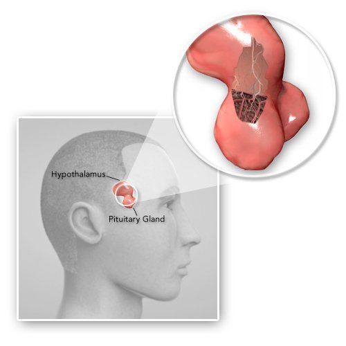
Specialized nerve cells descend down from the hypothalamus into the pituitary gland.

Illustration not drawn to scale.

Keeping the Signal Going

Sitting atop the kidneys, the adrenal glands receive nerve and chemical signals initiated by cells in the hypothalamus. Nerve signals activate the release of epinephrine into the bloodstream.

When chemical messengers arrive via the bloodstream, they dock on to receptors and begin a cell signaling cascade that results in the production of cortisol. Cortisol is released into the blood stream where it begins signaling cascades in several cell types, resulting in an increase in blood pressure, increase in blood sugar levels, and suppression of the immune system.

In Detail: The Cortisol Production Signaling Cascade
Pituitary Release

When nerve cells in the hypothalamus fire, corticotrope cells in the pituitary gland are stimulated to release ACTH, a chemical messenger, into the bloodstream.

Adrenal Gland

The ACTH chemical messengers, sent from the pituitary gland into the bloodstream, arrive at the adrenal cortical cells.

When the splanchnic nerve in the hypothalamus fires, a signal is sent down the spinal cord directly to the adrenal gland.

A Boost of Energy

Signaling molecules from several origins work to provide an energetic boost in a variety of ways. When epinephrine binds to receptors on liver cells, it triggers a signaling cascade that produces glucose from larger sugar molecules. Circulating cortisol sets fatty acids free to be transformed into energy. These molecules are rapidly excreted into the bloodstream, supplying a boost of readily available energy for muscles throughout the body, priming them for exertion.

In Detail: The Glycogenolysis Signaling Cascade
Epinephrine Release

The splanchnic nerve stimulates chromaffin cells in the adrenal gland to release epinephrine, a chemical messenger, into the bloodstream.

Proteins in Real Life

Often, it is necessary to take some artistic license when creating images of proteins and other molecules. So what do they really look like? Molecular models provide a glimpse into their tiny world and help us understand how they work. Take a look at some scientifically accurate models of the molecules and proteins from the movie.

Stylized phosphorylase kinase and activated glycogen phosphorylase (orange) fromThe Fight or Flight Response movie.

What phosphorylase kinase really looks like (purple), its lobes decorated with inactive glycogen phosphorylase (green).

Image courtesy Catherine Vénien-Bryan, PhD, University Research Lecturer, University of Oxford. Reproduced from Structure, 2002, Vol. 10, No. 1, 9. © 2002 with permission from Elsevier.

Proteins in real life

One Messenger, Many Reactions

Epinephrine is an important cell signaling molecule in the fight or flight response. Also known as adrenaline, epinephrine is an efficient messenger that signals many cell types throughout the body with many effects. In the lungs, epinephrine binds to receptors on smooth muscle cells wrapped around the bronchioles. This causes the muscles to relax, dilating the bronchioles and allowing more oxygen into the blood. At the sino-atrial node of the heart, epinephrine stimulates pace maker cells to beat faster. This increases the rate at which other chemical signals, glucose and oxygen are circulated to the cells that need them. Epinephrine also contracts specific types of muscle cells below the surface of the skin, causing beads of perspiration and raised hairs at the surface.

Epinephrine

Epinephrine, a chemical messenger.

A Complicated Orchestration

The fight or flight response is a complicated systemic reaction. These are just some of the instantaneous messengers and physiologic changes involved.

In fact, the initial perception of a threat or danger is also received by an area in the brain stem that begins yet another axis of communication and response involving the release of the messenger norepinephrine. Like cortisol and epinephrine, norepinephrine travels throughout the body, triggering cell signaling cascades in a number of cell types.

Regardless of their kind, or point of origin, cell signaling molecules involved in the fight or flight response work closely together. Their overall effect is an increase in circulation and energy to certain body systems and a downshift of less important ones into maintenance mode. In this way, the fight or flight response prepares the body for extreme action.

Heart
References

References

ACTH signal cascade:

Margioris, Andrew N., and Tsatsanis, Christos (2002, December 23). ACTH action on the adrenal. Retrieved September 10, 2008, from Endotext.org Web site:http://wwwotext.org/adrenal/adrenal5/adrenal5.htm

Jefcoate, Colin (2002). High-flux mitochondrial cholesterol trafficking, a specialized function of the adrenal cortex. Journal of Clinical Investigation, 110(7), 881-890.http://www.jci.org/articles/view/16771

Glycgenolysis signal cascade:

King, Michael W. (2000, April 12). Glycogen Metabolism. Retrieved September 10, 2008 from The THCME Medical Biochemistry Page Web site:http://dwb.unl.edu/teacher/nsf/c11/c11links/web.indstate.edu/thcme/mwking/glycogen.html

Phosphorylase kinse structure and function:

Benien-Bryan, Catherine (2002). Electron microscope studies. Retrieved September 10, 2008 from Oxford University, Laboratory of Molecular Biophysics Web site, Laboratory Journal 2002:http://biop.ox.ac.uk/www/lj2002/cvb/CVB_journal.html#figure3

Pacemaker heart cell description:

Richardson, Daniel R., Randall, David C., and Speck, Dexter F. (1998). Cardiopulmonary System (p. 94). Hayes Barton Press. http://books.google.com/books?id=D4GR3Znx8AEC&pg=PA94&lpg=PA94&dq=pacemaker+cell+round&source=web&ots=RpefTwL0Wn&sig=uMbYYVJdhSaiRZtNjleGzanq_5c


APA format:

Genetic Science Learning Center. (2020, July 31) How Cells Communicate During Fight or Flight. Retrieved March 09, 2024, from https://learn.genetics.utah.edu/content/cells/fight_flight/

CSE format:

How Cells Communicate During Fight or Flight [Internet]. Salt Lake City (UT): Genetic Science Learning Center; 2020 [cited 2024 Mar 9] Available from https://learn.genetics.utah.edu/content/cells/fight_flight/

Chicago format:

Genetic Science Learning Center. "How Cells Communicate During Fight or Flight." Learn.Genetics. July 31, 2020. Accessed March 9, 2024. https://learn.genetics.utah.edu/content/cells/fight_flight/.