Foundational Toolkit Genes

diverse eyes

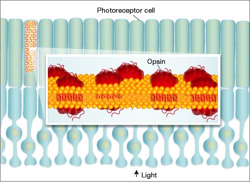
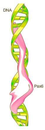
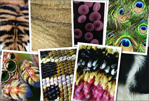
Given the variety of eyes found throughout the animal kingdom, evolutionary biologists once thought eyes had evolved independently dozens or even hundreds of times. Thanks to DNA sequencing and other molecular tools, we know today that modern eyes are built from many of the same genes. Ancient "toolkit" genes (such as opsins, Pax, and Otx) first evolved in a primitive ancestor that gave rise to all animals with eyes. These genes have been preserved throughout evolution, and today we still find them at work in all types of eyes. The diversity of modern animal eyes is the result of refinements and specializations built on top of this basic genetic framework.

Not all eye features are built using the same genes. Lenses, for example, are refinements that arose when different genes were recruited to perform a similar task in different organisms. Similarly, shielding pigments arose from a variety of genes.

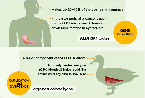
Molecular analysis has shown that many genes in the eye, whether shared among organisms or not, had other functions first. They were later recruited to take on second jobs.

All Eyes Use Opsins

Opsins detect light. Before being recruited into the eye, opsins had a variety of other jobs. In modern organisms that don't have eyes—such as archaea, fungi, green algae, protists, and simple animals—members of the opsin family act as ion pumps, sensory molecules, light-gated ion channels, and circadian-rhythm regulators.

Light-detecting opsins had to evolve only once. The thousand or more modern animal opsins are all modified versions of one that was present in a shared ancestor that lived more than 600 million years ago. Different opsins detect a variety of intensities and wavelengths of light. Many species have multiple opsins, allowing them to see a broad range of wavelengths, thus forming the basis of color vision. Each animal's opsins are tuned to the wavelengths of light that are most important to its lifestyle and environment.

Photoreceptors

Genes Are Universal Regulators

Pax 6

Defects in the Pax6 gene cause the loss of eye structures in humans, mice, and fruit flies. Mouse photos reprinted by permission from Macmillan Publishers Ltd: Nature 354(6354):522-5, ©1991. Human and fruit fly photos reprinted under Open Access License; Originally published in Washington, N. L. et al, PLoS One Biology 7(11): e1000247. doi:10.1371/journal.pbio.1000247

For eyes to function properly, many different proteins must interact. Pax proteins are regulators: they attach directly to the DNA and turn on the necessary genes in the right place at the right time. These regulators perform similar jobs in animals as diverse as insects and vertebrates, so they must have evolved before the two lineages split. In fact, nearly every animal that grows eyes does so with the help of Pax genes. Even organisms without eyes, such as sponges, have Pax genes; this finding suggests that Pax proteins originally controlled other processes and were later recruited into the eye.

In the mid-1990s, researchers were suprised to discover that fruit flies, mice, and humans who were born missing eye structures had defects in the same gene. This gene, called Pax6 (or eyeless in flies), is required for normal eye development in all animals with bilateral symmetry. Even in eyes that look very different, Pax6 functions in much the same way. When placed in a fly, the mouse Pax6 gene activates all the genes necessary to form a normal, functional fly eye (not a mouse eye).

Crystallins Have Diverse Origins

Tightly packed into a nearly crystal array, crystallin proteins make up the lens and cornea, the transparent light-gathering structures in many types of eyes. While all crystallins share many features—they're water soluble, transparent, and stable at high concentrations—they have an enormously diverse evolutionary history; different crystallins have evolved independently from a broad assortment of stress proteins and metabolic enzymes.

In some animals, crystallins participate in a type of job sharing: at low concentrations in other parts of the body, they still carry out their original stress or metabolic roles. In other animals, the original gene has been duplicated. One copy still does its old job while the other copy has been modified by natural selection into a high-performing lens protein. This theme of "duplication and divergence" is common throughout evolution.

Not only do diverse crystallins act alike, they are also controlled by the same regulatory genes:regulatory switches where Pax proteins can attach have evolved independently in front of unrelated crystallin genes.

Human and Duck comparison

Shielding Pigments Come in Many Forms

Scales

Many pigments that provide shielding in the eye also color other parts of the body. Starting at top left: tiger fur, elephant skin, red blood cells, peacock feathers, spider body, snake scales, moth scales, dog fur.

In simple eyes, dark pigments shield the photoreceptors, giving tiny, transparent animals more information about light direction. In more-complex eyes, pigments absorb light after it passes through photoreceptors, preventing it from scattering and degrading image quality, as well as from damaging sensitive tissues.

A wide variety of dark-colored molecules act as shielding pigments in different types of eyes, and many eyes contain more than one type of pigment. Common eye pigments include melanin (in vertebrates, cnidaria, planaria, and some nematodes), ommachromes (in molluscs and arthropods), pterins, guanidine, and hemoglobin.

As with crystallins, many genes involved in pigment production are also active in other parts of the body: melanins color skin, fur, and feathers; pterins color butterfly scales; ommachromes color spiders' bodies; hemoglobin transports oxygen in the blood.

References

References

References

Arendt, D., Hausen, H., Purschke, G. (2009). The 'division of labour' model of eye evolution.Philosophical Transactions of the Royal Society of London, Biological Sciences, 364(1531), 2809-2817. doi:10.1098/rstb.2009.0104

Lamb, T. D., Collin, S. P., Pugh, Jr., E. N. (2007).Evolution of the vertebrate eye: opsins, photoreceptors, retina and eye cup. Nature Reviews Neuroscience, 9(12), 960-976 (subscription required). doi:10.1038/nrn2283

National Library of Medicine & John Hopkins University (2001, April 3). MIM *100660 ALDEHYDE DEHYDROGENASE, FAMILY 3, SUBFAMILY A, MEMBER 1; ALDH3A1. Online Mendelian Inheritance in Man. Retrieved May 7, 2010, fromhttp://www.ncbi.nlm.nih.gov/omim/100660

Nilsson, D.-E. (2009). The evolution of eyes and visually guided behaviour.Philosophical Transactions of the Royal Society of London, Biological Sciences, 364(1531), 2833-2847. doi:10.1098/rstb.2009.0083

Shubin, N., Tabin, C., & Carroll, S. (2009).Deep homology and the origins of evolutionary novelty. Nature, 457, 818-823 (subscription required). doi:10.1038/nature07891

Tsai, M., Koo, J., & Howell, P. L. (2005).Recovery of argininosuccinate lyase activity in duck δ1 crystallin. Biochemistry 44(25), 9034-9044. doi:10.1021/bi050346s

Vopalensky, P. & Kozmik, Z. (2009). Eye evolution: common use and independent recruitment of genetic components.Philosophical Transactions of the Royal Society of London, Biological Sciences, 364(1531), 2819-2832. doi:10.1098/rstb.2009.0079

  • Funding

    Funding provided by grant 51006109 from the Howard Hughes Medical Institute, Precollege Science Education Initiative for Biomedical Research.


APA format:

Genetic Science Learning Center. (2014, December 1) Foundational Toolkit Genes. Retrieved April 15, 2024, from https://learn.genetics.utah.edu/content/senses/toolkit

CSE format:

Foundational Toolkit Genes [Internet]. Salt Lake City (UT): Genetic Science Learning Center; 2014 [cited 2024 Apr 15] Available from https://learn.genetics.utah.edu/content/senses/toolkit

Chicago format:

Genetic Science Learning Center. "Foundational Toolkit Genes." Learn.Genetics. December 1, 2014. Accessed April 15, 2024. https://learn.genetics.utah.edu/content/senses/toolkit.